Here at the UMD BIP lab, I have been looking for tracheal mites (Acarapis woodi) in honey bee samples. Tracheal mites live in the trachea of honey bees, they enter the tracheal tubes through the bee’s spiracles. Once inside the trachea, they puncture the wall of the trachea and feed on the bee’s haemolymph. An obvious sign that bees have a heavy tracheal mite infestation is when the trachea have brown/ dark red scarring. Cloudy trachea can also indicate the presence of mites. Clear trachea are USUALLY a sign of bees without tracheal mites. However, I have found mites in many bees that had clear tracheal tubes.
If you dissect 16 honey bees from a colony, and do not find mites in any of them, there is a 95% chance that tracheal mites are not present in the colony. I usually dissect 20 bees instead of 16, so that I have a few extra bees in case I accidentally cut the trachea out of the thorax of one of the bees.
Here is an outline of how I go about dissecting bees to look for tracheal mites.
- I take 20 bees from an alcohol sample of bees that was taken from an individual colony.
- Using a scalpel, with a No.10 surgical blade, I remove the bee’s abdomen
- Next, I put the bee on its back and cut off the head and first pair of legs.
- Then, I cut a thin cross section of the thorax and place it onto a microscope slide.
- I cut the first 10 bees and place them all on one slide. Then I place a small drop of 85% Lactic Acid onto each thoracic disk. The 85% Lactic Acid dissolves the bees muscle tissue and makes the trachea much easier to see.
- I repeat this process for the second set of 10 bees, and place them onto another slide. After applying the Lactic Acid, I usually wait about 15-25 minutes for the muscle tissue to dissolve/ lighten in color
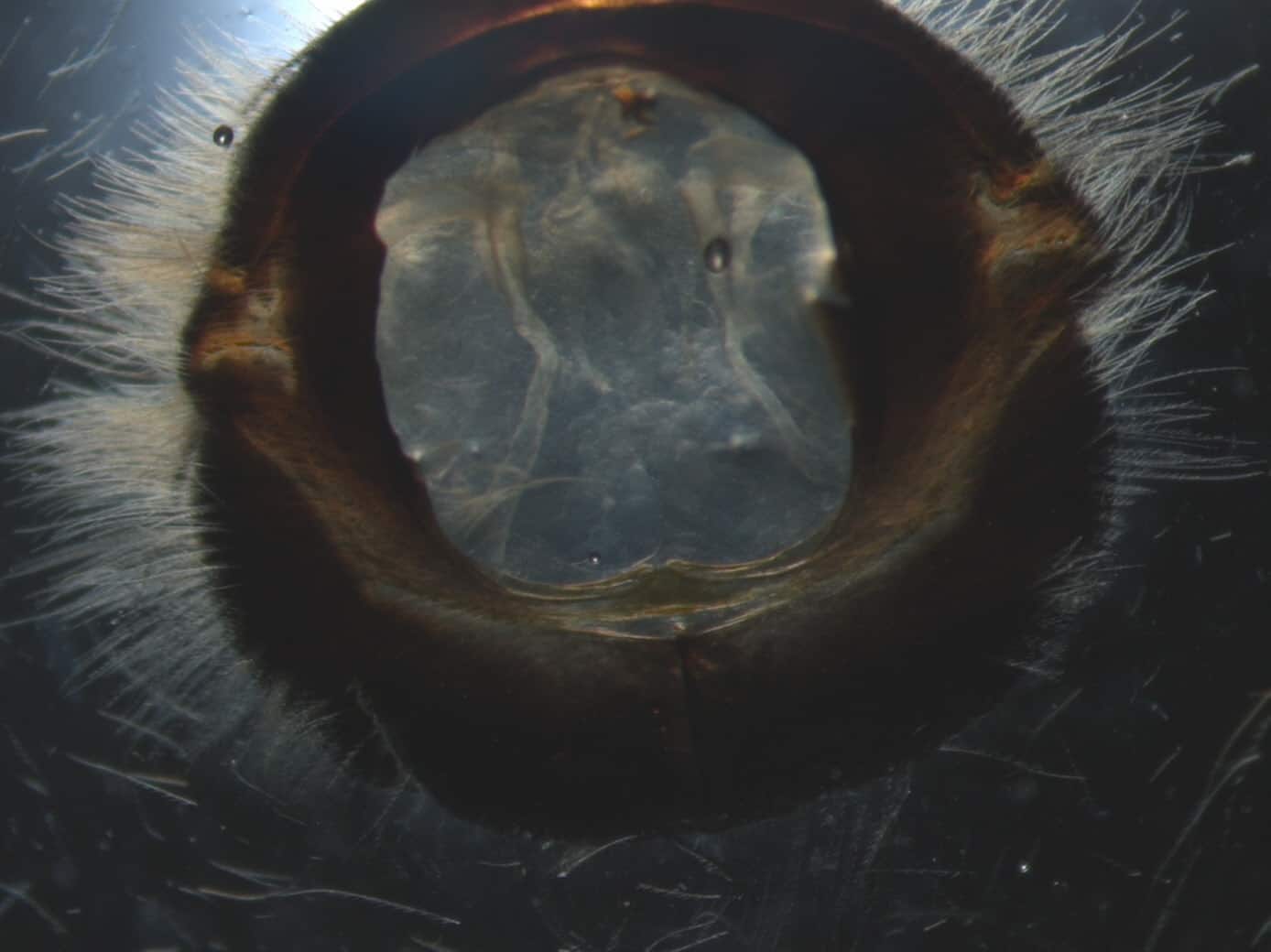
- Finally, I look at the slides under a dissecting microscope and scan over each bee to look for tracheal mites, or the scarring they make in the trachea.
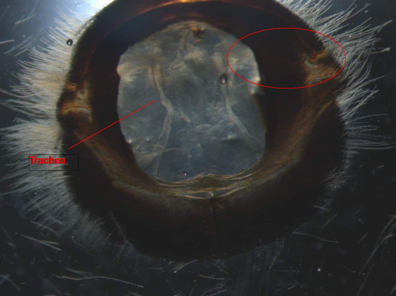
When looking at the thoracic disk from the top, the part of the trachea that is attached to the spiracle is obstructed from view. It is very important to look at this part of the trachea because mites enter the tracheal tubes through the spiracles. Bees with a low infestation are likely to have the mites close to the spiracle, because the mites have not moved further into the tube. Also, this area of the trachea is a bit wider, giving the mites more room. You can view this part of the tracheal tube by removing the collar of the bee, or by flipping the disk upside down and pulling out the tube for closer examination. I use #5/45 forceps with an angled tip to inspect the thoracic disks; you can use the forceps to dig through the muscle tissue to get a better look at the trachea.
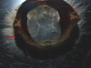
- The trachea is labeled on the left. The red circle on the right side of the image shows the location of the obstructed portion of trachea that attaches to the spiracle. The circled portion of the trachea is covered up by the “collar” which is a portion of the 1st thoracic segment of the bee (Prothorax).
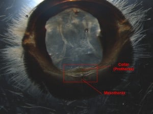
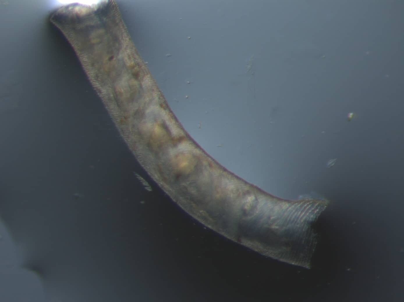
By pulling up on this section of the collar, you can remove it, and view the obstructed portion of the trachea.