This summer I have had the good fortune of continuing my work in the vanEnglesdorp lab, and I have learned a variety of new techniques for assessing hive health. The one I will be talking about today is a project I have just begun work on- bee autopsies. This may sound gruesome, and maybe it is, but I have thoroughly enjoyed learning how to correctly dissect and evaluate honey bee digestive tract health. This dissection, in the most basic terms, requires that the entire contents of the abdomen be removed and examined. This includes structures from the crop, or honey stomach, all the way to the sting sac.
For those of you without a background in entomology below are a few pictures of my dissections with labeled anatomy and a brief explanation of how these parts all function to keep the bee buzzing.
The Foregut
The foregut is composed of the pharynx, esophagus, and crop. The latter is the only part that is contained within the abdomen, and is often referred to as the “honey stomach.” This amazing little structure can hold up to 100 mg of nectar, although the usual load is only about 20-40 mg.1 For perspective, this means that a pound of nectar (notice this isn’t even the final honey product) takes about 12,000-24000 trips outside the hive to collect.1
The above picture is one of my early dissections, pictured with the bee thorax and head for perspective. Some of the main structures of the abdomen are highlighted and will be discussed presently.
The Midgut
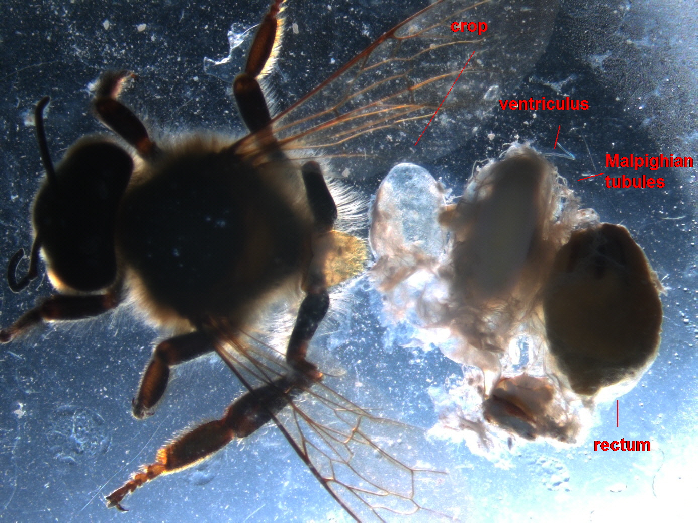
The midgut is comprised of the proventriculus, the ventriculus, and the small intestine. The proventriculus controls the flow of honey into the ventriculus, and also collects pollen in specialized pouches. These pouches allow pollen to be passed through the ventriculus tract as a single bolus.1 The ventriculus is lined with epithelial cells which constantly detach from the ventriculus lining to expel enzymes. These epithelia are constantly regenerated to keep the correct enzymes active in the ventriculus.1 Here, inside the ventriculus, is where the fungal parasite nosema “attacks.”1 If you haven’t been keeping up with our lab’s blogs, this is a parasite of great interest that will hopefully become a good indicator of hive health. From the ventriculus food is moved by peristalsis to the small intestine, an organ with pleated walls to increase nutrient absorption. Separating the large and small intestine is the pyloric valve. Another structure, technically not part of the GI tract connects here. This is the Malpighian tubules, which connect to the small intestine just before the pyloric valve. This structure is exceedingly important as these tubules essentially function as the kidneys of the honey bee. Like the human kidney they filter the bee’s circulatory fluid, which in this case is hemolymph. Unlike human kidneys, there are about 100 of these tubules which drain into the small intestine instead of a urinary tract.

The above picture was taken by Michael Andree, and shows the ventriculus, small intestine, and malpighian tubules that are reduced in number and size. This is one of the indicators of bee health assessed in the autopsy report.
The Hindgut
This section of the digestive tract is made up of only 2 structures, the rectum and anus. The rectal contents are mostly undigested pollen husks, pollen fat globules, and spent ventriculus epithelia.1 If bees have been fed food substitutes fermentation of the undigested products can cause an increase in hive temperature. In the winter this may even get extreme enough to cause premature brood laying.1

The above picture is was taken by Michael Andree, and shows 3 bee rectums that are at varying degrees of fullness. From left to right they may be described as thin, half full, and full. This is another dimension used in the autopsy report to assess bee health.
The poison sac and attached sting gland is situated in the posterior of the abdomen and is filled with the bee’s venom. This venom is mostly formic acid, hence the colloquial name for the sting gland, the acid gland. The inside of the gland has thick cuticular lamina which creates folds and rings to keep the transverse neck of the gland open so that the venom may be released.2

The above picture is one of the venom sac (left) and sting gland (right) taken by Michael Andree.
Autopsy Assessment
Now that you are familiar with the various abdomen structures of the honey bee, I will give a quick list of how each of these components is assessed.
General- Black tissue (present or absent), White nodules (present or absent. These may be attached to tergites, free floating in the abdomen, or attached to the GI tract)
Ventriculus- Coloration (light, dark, or very dark), Size (small, medium, or large)
Malpighian tubules- Color (normal, slightly discolored, very discolored), Number/Size (normal or reduced), Iridescence (normal- non-iridescent, abnormal- iridescent spots)
Pyloric Valve- Scarring (abnormal- scarred)
Rectal contents- Size (Full, half full or thin), Color (light, dark, very dark), Consistency (soft, semi-hard or hard), Food Packet presence (present or absent)
Venom Sac- Color (clear or discolored), Debris (present or absent)
Venom gland- Size (normal, slightly swollen, or swollen), Color (clear, slightly discolored, or very discolored), Melanosis (present or absent)
All of these components are to be analyzed in a set of bees that were sampled through the National Honey Bee Survey. The set of interest has had data collected about the pesticides used on plants these bees fed on. It is our hope that we may be able to eventually correlate honey bee health with the pesticides used in the area. In this way methods for honey bee preservation in the presence of pesticides can be developed.
I hope you have enjoyed learning a little bit about the squishy bits of bees. Don’t forget about your pollinators and above all, Bee Informed!
References
1 Dade, H. A. Anatomy and Dissection of the Honeybee. London: International Bee Research Association, 1977. Print.
2 Snodgrass, R. E. Anatomy of the Honey Bee. Ithaca, NY: Comstock Pub. Associates, 1956. Print.