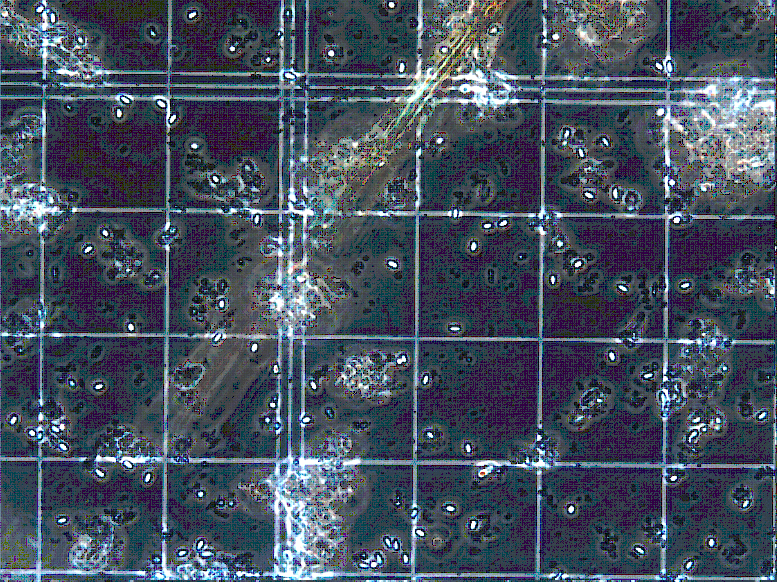
Looking into the eyepiece of my microscope at the water-mount slides I have prepared, hardly anything I see resembles honey bees. Coincidentally, everything appears a monochromatic amber-gold very close to the color scheme of the inside of a hive. But that is really where the similarity stops. Instead of seeing a humming colony of busy, hard-working bees, my eyes sift through a collection of stationary debris, microbes, and pollen.
I am Anna Wallis and I work with Heather Eversole in a lab processing samples taken from hives from all over the United States for honeybee diseases. I am a part of the fantastic team at the Bee Research Lab (BRL) in Beltsville, Maryland under Karen Rennich, who introduced Heather and me a few weeks ago.
What I am looking for under the microscope are Nosema spores. Nosema is a protozoan, a kind of single-celled organism related to fungus, that causes nosema disease in honey bees. The disease infects the ventriculus, or the midgut, in honey bees. It causes an obvious swelling and whitening of the digestive organ and includes symptoms like diarrhea, sometimes visible as yellow streaks on the outside of a hive. (Poor bees!)
Nosema is unable to be quickly identified by simply observing the bees and the hive. However, it is fairly easy for a trained eye to diagnose it in a lab with a microscope, by inspecting dissected bees or fecal matter. At BRL, we follow the procedure outlined in the Diagnosis of Honey Bee Diseases booklet by the USDA. Essentially, we take a sample of 100 bees and grind them to a pulp with a rolling pin in a plastic ziploc baggie and add water to release the contents of the bee’s ventriculus. For me, it feels like kind of a strange thing to do–grind up bees, that is–after spending so much time caring for hives this summer, painstakingly removing and replacing each box and frame trying not to pinch too many between the wood pieces or my hive tool. But, it only takes a very small sample from each hive and a couple of minutes at the microscope to find out this very important information.
Under the microscope, Nosema looks like shiny little ovals. We have also called them “cigar-” and “sesame-seed-shaped” here in the lab, to help first-timers find them more easily. They have perfectly smooth, well-defined edges, and they have a characteristic glow around their outer edges. It makes them seem very alien and other-worldly against the irregular bits of microscopic rubble around them.
It is never a good thing to find signs of a disease in a sample of bees, but I have to admit that I sometimes get a twinge of satisfaction when I find a spore–it is sort of like the gratification you get from picking out a four-leaf clover from the rest of the grass around it, like you are on a treasure hunt. Luckily, so far in the BIP survey Nosema spores have been few and far between. These bees appear to be some of the healthiest of all the bees that have come through our lab–which is great news since the beekeepers undergoing this extensive survey breed the queens that are used to populate nearly all the rest of the hives in the country!